Sep 15, 2008

ULNAR NERVE ANATOMY


The ulnar nerve derives from C8 and T1 nerve roots. It runs on the medial aspect of upper arm, and gives off no branches in the upper arm. It passes posterior to the medial epicondyle of the humerus to enter the cubital tunnel. Near elbow, ulnar nerve gives motor branches to flexor carpi and medial portion of flexor digitorum profundus.


In forearm, it gives rise to a palmar cutaneous branch which arises near the middle of the forearm and supplies the skin on the medial part of the palm, and the dorsal cutaneous branch which arises in the distal half of the forearm and supplies cutaneous sensation on the dorsal, ulnar surface of the hand and digits 4 and 5.


At the wrist, the nerve enters Guyon's canal and divides into a superficial sensory and deep motor branch. The superficial branch supplies sensation to the palmar surface of the ring and the little fingers. The deep motor branch supplies abductor digit minimi flexor digit minimi muscles, opponens digiti minimi, third and fourth lumbricals, the palmar and dorsal interossei, the flexor pollicis brevis and adductor pollicis brevis and first dorsal interosseous. In essence, ulnar nerve supplies all small muscles of hands except abductor pollicis brevis, flexor pollicis brevis, opponens pollicis and 1st and 2nd lumbricals (which are supplied by median nerve).

Reference:
  1. Richard S Snell, Clinical Anatomy: Lippincott Williams & Wilkins, 7th edition
  2. Preston DC. Distal Median Neuropathies. In: Entrapment and other focal neuropathies; Neurologic Clinics: WB Saunders company, August 1999
  3. http://depts.washington.edu/anesth/regional/ulnarnerve.html

SENSORY CONDUCTION STUDIES - PARAMETERS


Sensory axons are evaluated by stimulating a nerve while recording the transmitted potential (known as sensory nerve action potential or SNAP) from the same nerve at a different site. (See orthodromic and antidromic studies)
SNAPs are of much lower amplitude (measured in millivolts) than compound muscle action potentials, and they often require averaging of multiple responses. Three main parameters recorded with sensory nerve conduction studies are – latencies, amplitudes and conduction velocity.


LATENCY

Latencies reflect time taken (in milliseconds) for an impulse to travel from the point of stimulation to the recording electrode. Two types of sensory distal latencies are used – peak latency and onset latency.

SNAP AMPLITUDE
This is a semiquantitative measure of the number of sensory axons that conduct between the stimulation and recording sites. It is expressed in microvolts.

CONDUCTION VELOCITY
This requires stimulation at a single site only because unlike CMAP, SNAP is true nerve action potential. (See motor conduction studies – parameters).


Reference:


  1. Aminoff, MJ. Electrodiagnosis in Clinical Neurology: Nerve conduction studies, New York: Churchill Livingston, 4th edition
  2. Kimura J. Electrodiagnosis in disease of nerve and muscle: Principles and Practice, New York: Oxford V. Press, 3rd edition


MOTOR CONDUCTION STUDIES - PARAMETERS


When a motor or mixed nerve is stimulated and recording is made by placing electrodes over a muscle supplied by that nerve, the recorded potential is known as compound muscle action potential or CMAP. There are three main parameters of CMAP, which are routinely evaluated during motor nerve conduction studies. They are latency, amplitude and conduction velocity.

LATENCY
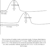
This is the time in milliseconds between nerve stimulation and initial deflection from baseline. It reflects the time required for action potential to travel along the fastest-conducting axons to activate the muscle fibers.


  • The latency includes not only the time taken for impulse to travel along the nerve till it reaches nerve terminal, but also the time taken for neuromuscular junction transmission and muscle activation.
  • Whenever possible, the nerve is stimulated at two points: a distal point near the recording site (distal latency) and a more proximal point (proximal latency).
  • Prolonged latencies are usually taken as evidence of demyelination.


AMPLITUDE
This is usually measured as height in millivolts of CMAP, from the baseline to the negative peak.

  • CMAP amplitude is a semiquantitative measure of the number of axons conducting between the stimulating and the recording points.
  • Decreased CMAP amplitudes usually suggest either axon loss or conduction block from demyelination located b/w the stimulation site and recorded muscle. But it can be due to reasons other than motor nerve dysfunction (e.g. neuromuscular junction, muscle fiber etc).


CONDUCTION VELOCITY

Measurement of differences in distance and latency b/w proximal and distal stimulation sites allows calculation of conduction velocity in the segment of nerve b/w the site of stimulation and is expressed in meters per second.

  • Normal conduction velocities are from 40-50 m/sec in the legs and from 50-70 m/sec in the arms.
  • Motor conduction velocity can not be calculated by performing a single stimulation. This is because, the latency of compound muscle action potential reflects transmission across nerve, junction and muscle, measurement of true conduction velocity across the nerve will necessarily require stimulation at two points.
  • Decrease in conduction velocities is usually taken as sign of demyelination.

Reference:

  1. Kimura J. Electrodiagnosis in disease of nerve and muscle: Principles and Practice, New York: Oxford V. Press, 3rd edition
  2. Preston DC, Shapiro BE. Electromyography and Neuromuscular Disorders, Boston: Butterworth-Heinemann


RECORDING ELECTRODES - FOR SENSORY STUDIES


The arrangement of active (G1) electrode, reference electrode (G2), and ground electrodes follows the same principle as in motor conduction, except that, in place of muscle, G1 and G2 electrodes are placed over the sensory nerves.


Sensory nerves are evaluated by stimulating a nerve while recording from the same nerve at a different site.


Antidromic sensory NCSs are performed by stimulating nerve proximally and recording distally along the sensory nerves, whereas orthodromic studies are obtained by stimulating it distally and recording it proximally along the nerve (i.e. recording sensory potentials as they travel towards CNS). See figure.

Ring electrodes are convenient to record the sensory potentials from digital nerves over the proximal and distal interphalangeal joints.


Reference:
  1. Sethi RK, Thompson LL. The Electromyographer’s Handbook, Boston/Toronto: Little, Brown and Company, 2nd edition
  2. Aminoff, MJ. Electrodiagnosis in Clinical Neurology: Nerve conduction studies, New York: Churchill Livingston, 4th edition


RECORDING ELECTRODES – FOR MOTOR STUDIES


Motor NCSs are performed by stimulating a motor or mixed peripheral nerve while recording the CMAP from a muscle innervated by that nerve. Standard anatomic locations of recording electrodes (and stimulation) provide reproducible potentials.


For motor nerve conduction studies, the preferred method is belly-tendon recording. In this arrangement, active electrode (known as G1) is placed on the belly of the muscle and reference electrode (known as G2) on the tendon. The ground electrode is usually placed between the stimulating and recording electrodes.


The nerve is stimulated at two or more points along its course. Typically, it is stimulated distally near the recording electrode and more proximally to evaluate its proximal segment. This is important, since for measurement of conduction velocity in motor conduction studies, single site stimulation may not be enough (see later).


Reference:

  1. Sethi RK, Thompson LL. The Electromyographer’s Handbook, Boston/Toronto: Little, Brown and Company, 2nd edition
  2. Aminoff, MJ. Electrodiagnosis in Clinical Neurology: Nerve conduction studies, New York: Churchill Livingston, 4th edition

RECORDING PROCEDURE - STIMULATING NERVES


Nerve stimulation is achieved with surface electrodes placed over a nerve where it is relatively superficial, such as ulnar nerve at elbow. The stimulator has a cathode and an anode. Normally both electrodes are placed over the nerve trunk, with the Cathode being Closer to the recording site (SEE INTRODUCTION).


Supramaximal stimulation of a nerve is required. This is to make sure that all axons have been depolarized. To achieve supramaximal stimulation, a gradually increasing stimulus current is applied, resulting in progressive increase in the size of the compound muscle action potential as more and more axons in the nerve are activated. When stage is reached, where no further increase in amplitude is seen, current is increased by additional 20-30% to ensure that no further change in amplitude occurs.


Reference:
1. Aminoff, MJ. Electrodiagnosis in Clinical Neurology: Nerve conduction studies, New York: Churchill Livingston, 4th edition
2. Kimura J. Electrodiagnosis in disease of nerve and muscle: Principles and Practice, New York: Oxford V. Press, 3rd edition